I. Learning Objectives:
- Identify the parts of the heart and tell each functions.
- Draw and label the parts of the heart
- Show proper care and concern about the healthiness of the heart.
II. Concept/Terminologies:
1. Heart – one of the most important organs in the entire human body.
a. atrium- receives blood.
b. ventricle-sends blood.
III. References:
1. Science for Daily Use VI, pp. 10-12
2. Into the Future Science & Health 6, pp. 6-9 IV.
IV.Review of Related Lesson:
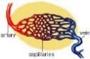
1. What are the major parts/organs of circulatory system.
Identify in the picture

V. Lecture Presentation:
- Motivational Question:
Feel your heart. What does your heart do for you? What do you think is the size of the heart?
2. Presentation:
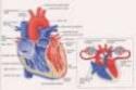
What are the parts of the heart?
See Picture:
Refer to pages 6-9, Into the Future Science & Health 6.


Establishment of the four-chambered heart, along with the pulmonary and systemic circuits, completely separates oxygenated from deoxygenated blood. This allows higher the metabolic rates needed by warm-blooded birds and mammals. The above image is from http://www.biosci.uga.edu/almanac/bio_104/notes/may_7.html. The human heart is a two-sided, 4 chambered structure with muscular walls. An atrioventricular (AV) valve separates each auricle from ventricle. A semilunar (also known as arterial) valve separates each ventricle from its connecting artery. The heart beats or contracts 70 times per minute. The human heart will undergo over 3 billion contraction cycles during a normal lifetime. The cardiac cycle consists of two parts: systole (contraction of the heart muscle) and diastole (relaxation of the heart muscle). Atria contract while ventricles relax. The pulse is a wave of contraction transmitted along the arteries. Valves in the heart open and close during the cardiac cycle. Heart muscle contraction is due to the presence of nodal tissue in two regions of the heart. The SA node (sinoatrial node) initiates heartbeat. The AV node (atrioventricular node) causes ventricles to contract. The AV node is sometimes called the pacemaker since it keeps heartbeat regular. Heartbeat is also controlled by the autonomic nervous system. The cardiac cycle. Image from Purves et al., Life: The Science of Biology, 4th Edition, by Sinauer Associates (www.sinauer.com) and WH Freeman (www.whfreeman.com), used with permission. Blood flows through the heart from veins to atria to ventricles out by arteries. Heart valves limit flow to a single direction. One heartbeat, or cardiac cycle, includes atrial contraction and relaxation, ventricular contraction and relaxation, and a short pause. Normal cardiac cycles (at rest) take 0.8 seconds. Blood from the body flows into the vena cava, which empties into the right atrium. At the same time, oxygenated blood from the lungs flows from the pulmonary vein into the left atrium. The muscles of both atria contract, forcing blood downward through each AV valve into each ventricle. Diastole is the filling of the ventricles with blood. Ventricular systole opens the SL valves, forcing blood out of the ventricles through the pulmonary artery or aorta. The sound of the heart contracting and the valves opening and closing produces a characteristic "lub-dub" sound. Lub is associated with closure of the AV valves, dub is the closing of the SL valves. Human heartbeats originate from the sinoatrial node (SA node) near the right atrium. Modified muscle cells contract, sending a signal to other muscle cells in the heart to contract. The signal spreads to the atrioventricular node (AV node). Signals carried from the AV node, slightly delayed, through bundle of His fibers and Purkinjie fibers cause the ventricles to contract simultaneously. The contraction of the heart and the action of the nerve nodes located on the heart. Images from Purves et al., Life: The Science of Biology, 4th Edition, by Sinauer Associates (www.sinauer.com) and WH Freeman (www.whfreeman.com), used with permission. An electrocardiogram (ECG) measures changes in electrical potential across the heart, and can detect the contraction pulses that pass over the surface of the heart. There are three slow, negative changes, known as P, R, and T. Positive deflections are the Q and S waves. The P wave represents the contraction impulse of the atria, the T wave the ventricular contraction. ECGs are useful in diagnosing heart abnormalities. Normal cardiac pattern (top) and some abnormal patterns (bottom). Images from Purves et al., Life: The Science of Biology, 4th Edition, by Sinauer Associates (www.sinauer.com) and WH Freeman (www.whfreeman.com), used with permission.
The Human Heart
 The heart is one of the most important organs in the entire human body. It is really nothing more than a pump, composed of muscle which pumps blood throughout the body, beating approximately 72 times per minute of our lives. The heart pumps the blood, which carries all the vital materials which help our bodies function and removes the waste products that we do not need. For example, the brain requires oxygen and glucose, which, if not received continuously, will cause it to loose consciousness. Muscles need oxygen, glucose and amino acids, as well as the proper ratio of sodium, calcium and potassium salts in order to contract normally. The glands need sufficient supplies of raw materials from which to manufacture the specific secretions. If the heart ever ceases to pump blood the body begins to shut down and after a very short period of time will die.
The heart is essentially a muscle(a little larger than the fist). Like any other muscle in the human body, it contracts and expands. Unlike skeletal muscles, however, the heart works on the "All -or-Nothing Law". That is, each time the heart contracts it does so with all its force. In skeletal muscles, the principle of "gradation" is present. The pumping of the heart is called the Cardiac Cycle, which occurs about 72 times per minute. This means that each cycle lasts about eight-tenths of a second. During this cycle the entire heart actually rests for about four-tenths of a second.
Make-up of the Heart.
 The walls of the heart are made up of three layers, while the cavity is divided into four parts. There are two upper chambers, called the right and left atria, and two lower chambers, called the right and left ventricles. The Right Atrium, as it is called, receives blood from the upper and lower body through the superior vena cava and the inferior vena cava, respectively, and from the heart muscle itself through the coronary sinus. The right atrium is the larger of the two atria, having very thin walls. The right atrium opens into the right ventricle through the right atrioventicular valve(tricuspid), which only allows the blood to flow from the atria into the ventricle, but not in the reverse direction. The right ventricle pumps the blood to the lungs to be reoxygenated. The left atrium receives blood from the lungs via the four pulmonary veins. It is smaller than the right atrium, but has thicker walls. The valve between the left atrium and the left ventricle, the left atrioventicular valve(bicuspid), is smaller than the tricuspid. It opens into the left ventricle and again is a one way valve. The left ventricle pumps the blood throughout the body. It is the Aorta, the largest artery in the body, which originates from the left ventricle.
 The Heart works as a pump moving blood around in our bodies to nourish every cell. Used blood, that is blood that has already been to the cells and has given up its nutrients to them, is drawn from the body by the right half of the heart, and then sent to the lungs to be reoxygenated. Blood that has been reoxygenated by the lungs is drawn into the left side of the heart and then pumped into the blood stream. It is the atria that draw the blood from the lungs and body, and the ventricles that pump it to the lungs and body. The output of each ventricle per beat is about 70 ml, or about 2 tablespoons. In a trained athlete this amount is about double. With the average heart rate of 72 beats per minute the heart will pump about 5 litres per ventricle, or about 10 litres total per minute. This is called the cardiac output. In a trained athlete the total cardiac output is about 20 litres. If we multiply the normal, non-athlete output by the average age of 70 years, we see that the cardiac output of the average human heart over a life time would be about 1 million litres, or about 250,000 gallons(US)! 3.
3. Summary:
a. What kind of organ is the heart?
b. What are the inner parts of the heart?
c. Describe the parts and function of each heart
d. Why is the ventricle thicker than the atrium?
e. What does the heart pump? Why is it important?
f. Can you live without the heart? Why?
g. What are thing you do in order to show your love and care in your heart?
VI. Learning Activities:
For more learning activities log on to www.pbs.org/wgbh/nova/eheart/human.html and www.pbs.org/wgbh/nova/heart/heartfacts/html
VII. Evaluation: Log on to www.prongo.com/quizstation
VIII. Assignment: Draw a heart and label it.


